MARINE MAMMAL RESEARCH UNIT - UBC

You can’t beat a healthy heart

You can’t beat a healthy heart
Marine Mammal Research Unit

New research is shedding light on the hearts of healthy marine mammals, and how they compare to human hearts

Everyone knows that heart health is important to human health. However, relatively little is known about the hearts of marine mammals, and how they compare to our hearts and those of other terrestrial mammals.
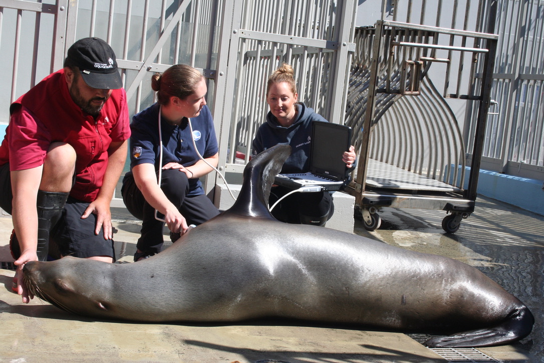
During a training session at the Vancouver Aquarium, Rhea Storlund holds an ultrasound probe to the chest of a Steller sea lion.

For marine mammals, the heart has to perform some extraordinary tricks. When marine mammals dive, their heart rates decrease to very low levels to allow the limited oxygen stored in their blood to be directed to where it is needed most.

Despite the critical role that the heart plays during diving, many questions remain about heart function in marine mammals. Even some of the most basic measurements of heart health that are routinely measured in people—such as heart rate and cardiac electrical activity—can be quite challenging to measure in marine mammals. Yet this information is critical for understanding how the hearts of marine mammals differ from terrestrial mammals. It is also needed to assess the cardiac health of individual animals.

An echocardiogram of a Steller sea lion heart showing the right ventricle (RV), left ventricle (LV), aortic root (Ao), and left atrium (LA).

Rhea Storlund, a PhD Candidate with the Marine Mammal Research Unit, brought together a team of veterinarians, animal health technicians, trainers, and researchers to examine the beating hearts of three species of pinnipeds at the Vancouver Aquarium. She and her team attached electrodes to a walrus, 5 fur seals and 8 sea lions to record electrocardiograms (ECGs), and also used an ultrasound to take picture of the left sides of the sea lions’ hearts.

The Pinniped Cardio Team.

The team shared their findings in a pair of recent papers. The first paper, published in the Journal of Zoo and Wildlife Medicine, provides insight into pinniped heart health. Using echocardiography—imaging the heart using ultrasound—the team was able to view, measure, and describe the left ventricle of Steller sea lion hearts. They also used electrocardiography to study the electrical signals that coordinate heartbeats in the same Steller sea lions, as well as in the northern fur seals and walrus. The values they reported are useful reference points for assessing cardiac health in pinnipeds under human care.

In a second paper—published in Frontiers in Physiology—Storlund and her colleagues went one step further by comparing the timing and coordination of cardiac muscle contractions between marine and terrestrial mammals. Because marine mammals have cardiovascular adaptations for diving, Storlund wondered if there were differences between the electrical activity of marine and terrestrial mammal hearts? This question formed the idea for this second paper.

Figures showing the relationships between body mass, ecological group (marine or terrestrial) and P wave duration (A), QRS complex duration (B), and QT interval duration (C) in mammals. All three ECG parameters increased with body mass, but the relationships were different for marine mammals (shown in blue) and terrestrial mammals (shown in green).

Storlund and her team compared the ECG values of 69 species of mammals (including the values from the walrus, Steller sea lions, and northern fur seals), and were surprised to discover that the heart rates of similar sized terrestrial and marine mammals were almost the same—despite the greater physiological challenges marine mammals incur by living in water. However, a closer inspection of the characteristic spikes of the ECG’s that make up individual heartbeats revealed several differences in how the muscles of their hearts contract. Storlund suspects that the differences they found between marine and terrestrial mammals are due to the unique size and shape of marine mammal hearts, which are flattened and have thicker walls compared to those of terrestrial mammals.

The work that Storlund led contributes significantly to identifying abnormalities and adaptations in the hearts of marine mammals. However, much more remains to be learned as Storlund and her team continues to press on with their cardiac research—one heartbeat at a time.


Rhea Storlund is a PhD candidate at the Marine Mammal Research Unit at the University of British Columbia

PublicationsPublication


2021
 
Cardiac examinations of anesthetized Steller sea lions (Eumetopias jubatus), northern fur seals (Callorhinus ursinus), and a walrus (Odobenus rosmarus).
Storlund, R.L., D.A.S. Rosen, M. Margiocco, M. Haulena and A.W. Trites. 2021.
Journal of Zoo and Wildlife Medicine 52(2):507-519.
abstract
Pinniped hearts have been well described via dissection, but in vivo measurements of cardiac structure, function, and electrophysiology are lacking. Electrocardiograms (ECGs) were recorded under anesthesia from 8 Steller sea lions (Eumetopias jubatus), 5 northern fur seals (Callorhinus ursinus), and 1 walrus (Odobenus rosmarus) to investigate cardiac electrophysiology in pinnipeds. In addition, echocardiograms were performed on all 8 anesthetized Steller sea lions to evaluate in vivo cardiac structure and function. Measured and calculated ECG parameters included P‑wave, PQ, QRS, and QT interval durations, P‑, R‑, and T‑wave amplitudes, P‑ and T‑wave polarities, and the mean electrical axis (MEA). Measured and calculated echocardiographic parameters included left ventricular internal diameter, interventricular septum thickness, and left ventricular posterior wall thickness in systole and diastole (using M-mode), left atrium and aortic root dimensions (using 2D), and maximum aortic and pulmonary flow velocities (using pulsed wave spectral Doppler). ECG measurements were similar to those reported for other pinniped species, but there was considerable variation in the MEAs of Steller sea lions and northern fur seals. Echocardiographic measurements were similar to those reported for southern sea lions (Otaria flavenscens), including 5 out of 8 Steller sea lions having a left atrial to aortic root ratio < 1, which may indicate that they have an enlarged aortic root compared to awake terrestrial mammals. Isoflurane anesthesia likely affected some of the measurements as evidenced by the reduced fractional shortening found in Steller sea lions compared to awake terrestrial mammals. The values reported are useful reference points for assessing cardiac health in pinnipeds under human care.
show/hide abstract View Reference


2021
 
Electrocardiographic scaling reveals differences in electrocardiogram interval durations between marine and terrestrial mammals.
Storlund, R.L., D.A.S. Rosen and A.W. Trites. 2021.
Frontiers in Physiology 12:690029
abstract
Although the ability of marine mammals to lower heart rates for extended periods when diving is well documented, it is unclear whether marine mammals have electrophysiological adaptations that extend beyond overall bradycardia. We analyzed electrocardiographic data from 50 species of terrestrial mammals and 19 species of marine mammals to determine whether the electrical activity of the heart differs between these two groups of mammals. We also tested whether physiological state (i.e., anesthetized or conscious) affects electrocardiogram (ECG) parameters. Analyses of ECG waveform morphology (heart rate, P-wave duration, and PQ, PR, QRS, and QT intervals) revealed allometric relationships between body mass and all ECG intervals (as well as heart rate) for both groups of mammals and specific differences in ECG parameters between marine mammals and their terrestrial counterparts. Model outputs indicated that marine mammals had 19% longer P-waves, 24% longer QRS intervals, and 21% shorter QT intervals. In other words, marine mammals had slower atrial and ventricular depolarization, and faster ventricular repolarization than terrestrial mammals. Heart rates and PR intervals were not significantly different between marine and terrestrial mammals, and physiological state did not significantly affect any ECG parameter. On average, ECG interval durations of marine and terrestrial mammals scaled with body mass to the power of 0.21 (range: 0.19 - 0.23) rather than the expected 0.25—while heart rate scaled with body mass to the power of -0.22 and was greater than the widely accepted -0.25 derived from fractal geometry. Our findings show clear differences between the hearts of terrestrial and marine mammals in terms of cardiac timing that extend beyond diving bradycardia. They also highlight the importance of considering special adaptations (such as breath-hold diving) when analyzing allometric relationships.

keywords     ECG, electrocardiogram, marine mammal, heart rate, anesthesia, allometry, cardiac timing, comparative electrophysiology
show/hide abstract View Reference